45 onion cells under microscope with labels
Microscopes and Cells - Biology I: Introduction to Cell ... Be sure to label the chloroplasts, the cell membrane, and the cell wall. Procedure 2. Onion Epithelial Cells. Obtain a new slide and use one half of the slide to examine onion cells. Cut a small piece of onion and break it by bending it in half. Using forceps, peel the thin membrane from the outer surface of a layer of onion. bmcbiol.biomedcentral.com › articles › 10Structure and function of mitochondrial membrane protein ... Oct 29, 2015 · Mitochondria can be seen in the light microscope, but their detailed internal structure is only revealed by electron microscopy. In the 1990s, the structure of mitochondria was investigated by electron tomography of thin plastic sections . While this yielded striking three-dimensional (3D) images of their internal membrane system, molecular ...
khabarban.com › a › 34719343یادواره سرلشگر شهید «محمدحسن قاسمی طوسی » برگزار می شود - مهر عبدالرضا بابایی عصر سه شنبه در نشست خبری شانزدهمین یادواره سرلشگر شهید محمد حسن قاسمی طوسی و 436 شهید شهرستان و نکوداشت روز نکا گفت، آنچه که انقلاب اسلامی را به پیروزی رساند جهاد و توجه ویژه به این مقوله بود که در واقع ...

Onion cells under microscope with labels
Cheek Cells Under The Microscope - YouTube Human cheek cells are made of simple squamous epithelial cells, which are flat cells with a round visible nucleus that cover the inside lining of the cheek.C... ocr.org.uk › Images › 643844-question-paper-depth-inOxford Cambridge and RSA Friday 16 October 2020 – Morning 1 (a) A student was observing onion epithelial cells using a light microscope. They photographed these cells and the image obtained is shown in Fig. 1.1. The student then made a drawing of a few cells from this image. The drawing is shown in Fig. 1.2. Fig. 1.1 cytoplasm cell wall large permanent vacuole ribosome Fig. 1.2 Required practical - using a light microscope - Cells in ... to show the outline of individual cells that make up the tissue, if the tissue is uniform A high power diagram is then produced - a detailed image of a part of the slide. It is usually drawn to...
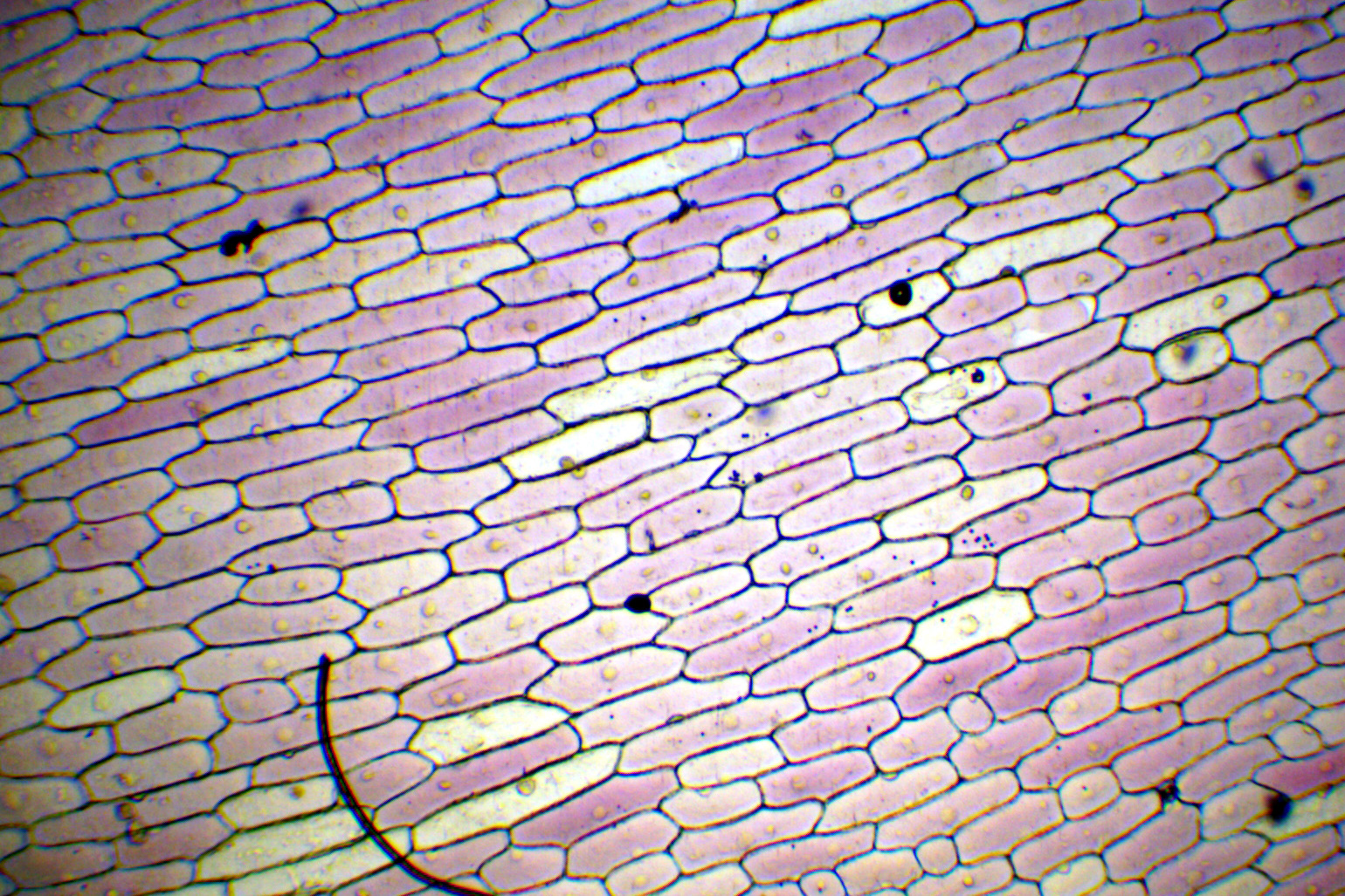
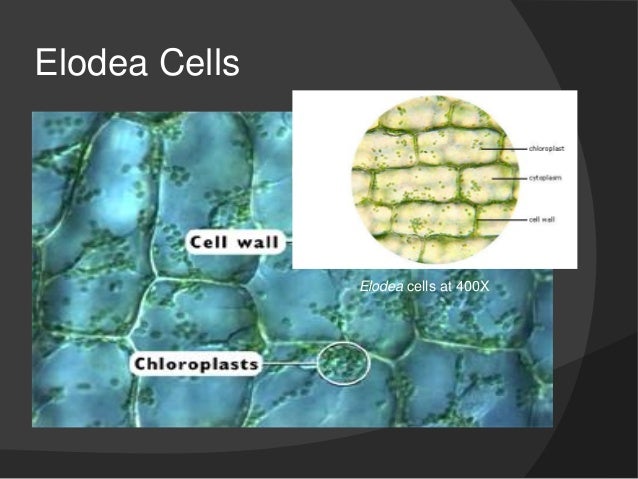
Onion cells under microscope with labels. The Microscope and Cells | Biology I Laboratory Manual Part 1: Microscope Parts . The compound microscope is a precision instrument. Treat it with respect. When carrying it, always use two hands, one on the base and one on the neck.. The microscope consists of a stand (base + neck), on which is mounted the stage (for holding microscope slides) and lenses. The lens that you look through is the ocular (paired in binocular scopes); the lens that ... Onion Skin Cells - Investigation - Exploring Nature 5. Observe the onion tissue under the microscope at 4x, 10x and 40x with lots of light (open diaphragm). Then slowly close the diaphragm while observing the image to find the best light for seeing cellular details. 6. Draw a section of onion skin cells at 10x magnification. Then switch to 40x and draw one cell and label it. Microscope Imaging Station. Gallery. - Exploratorium Chloroplasts and mitochondria move within Elodea leaf cells; nuclei are also visible as clear, fried-egg-shaped structures. Elodea are common freshwater aquarium plants. An elodea leaf was mounted in pondwater between a slide and coverslip with a silicon spacer. Images were taken on an inverted compound microscope using a 40x DIC objective and ... Blog, She Wrote - Embracing the Independent & Authentic ... Blog, She Wrote - Embracing the Independent & Authentic ...
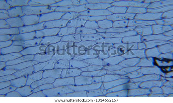
› bitesize › articlesCells and Reproduction - BBC Bitesize Onion cells are easy to see using a light microscope. ... A small tube placed under the skin of the upper arm. ... Five small tubes with labels and stoppers or lids Cress seeds Labels Cotton wool ... Observing Onion Cells under a Microscope - Blog, She Wrote you'll need to stain the onion cells before you observe them under the microscope. There are different types of stains depending on what type of cell you are going to look at. Iodine - dark stain that colors starches in cells. In an onion cell, it will make the cell wall more visible. It provides some contrast for viewing under a microscope. Microscopy Practical (Onion Cells) | Teaching Resources Presentation and practical handout for observing onion cells under a light microscope for teaching and revision. A step by step visual guide for all abilities. Can be used as a distance based learning tool during local covid lockdown and in classes where practicals are on-hold due to coronavirus. Content covered: Light microscope parts The following diagram shows cells of onion peel label ... In order to label them, we need to understand its anatomy and know about various structures present in it. Onion peel is an example of a plant cell whereas a human cheek cell is an example of an animal cell. Complete answer:
Observing Onion Cells Under The Microscope How to observe onion cells under a microscope. Onions are composed of several layers separated by thin membranes. In this activity, we will be using these thin membranes to observe onion cells in great detail. Obtaining a thin membrane from a bulb of onion is easy with the use of a pair of tweezers. For best results, use small, firm onions. Leaf Structure Under the Microscope When viewed under the microscope, it's possible to see the epidermal cells that tend to be irregular. In addition to the epidermal cells, one will also see the leaf spores (stomata) in between the epidermal cells. Typically, the stomata are bean shaped and will appear denser (darker) under the microscope. Biology Experiment Examination of Onion Cell in Light ... Place the single layer of onion cell epithelium on a glass slide. Make sure that you do not fold it over or wrinkle it. Place a drop of iodine stain on your onion tissue. Put the cover slip on the stained tissue and gently tap out any air bubbles. Observe the cells under 4x, 10x, and 40x with the diaphragm wide open. jnanobiotechnology.biomedcentral.com › articles › 10Nano based drug delivery systems: recent developments and ... Sep 19, 2018 · Nanomedicine and nano delivery systems are a relatively new but rapidly developing science where materials in the nanoscale range are employed to serve as means of diagnostic tools or to deliver therapeutic agents to specific targeted sites in a controlled manner. Nanotechnology offers multiple benefits in treating chronic human diseases by site-specific, and target-oriented delivery of ...
Microscopy
Microscope Cell Lab: Cheek, Onion, Zebrina - SchoolWorkHelper Microscope Cell Lab: Cheek, Onion, Zebrina Introduction The purpose of this lab was to use the microscope and identify cells such as animal cells and plant cells. This subject is important because in Biology, we will be using the microscope many times during different laboratory exercises.

Labeled Onion Cell Under Microscope 40x - Micropedia
Onion Cells Microscope High Resolution Stock Photography ... Onion cells under the microscope. Garden onion, Bulb Onion, Common Onion (Allium cepa), cell tissue of a garden onion with dyed chromosomes, light microscopy, x 120, Germany. Onion Cells under the Microscope. Onion skin cells under the microscope, horizontal field of view is about 0.61 mm. Detailed view of the cells of a red onion as seen ...
Cell lab
Animal Cell Under Light Microscope Labelled : Draw and ... Onion cell diagram labeled structure of animal cell and plant cell under microscope. An organelle found in large numbers in most cells, in which the biochemical processes of respiration and energy production occur. Under a light microscope, the cell membrane, nucleus and cytoplasm of a cheek cell (animal cell) can be observed.
)
Onion Cells Under Microscope Stock Footage Video 4946132 | Shutterstock
PDF Onion Cell Lab - SomeWaresInMaine Research Biology Onion Cell Lab page 1 of 3 Onion Cell Lab After you have completed the rest of this lab come back to this cover page DRAW & LABEL AN ONION CELL WITH ALL THE PARTS / ORGANELLES YOU OBSERVE UNDER 40X. Purpose: To observe and identify major plant cell structures and to relate the structure of the cell to its function.

Eugene PARK's Fantastic Microworld: Onion cells and yeasts
Looking at the Structure of Cells in the Microscope ... Both types of light microscopy are widely used to visualize living cells. Figure 9-7 Two ways to obtain contrast in light microscopy. (A) The stained portions of the cell reduce the amplitude of light waves of particular wavelengths passing through them. A colored image of the cell is thereby obtained that is visible in the ordinary way. (more...)

The Microscope
› 36111379(PDF) DiFiore's Atlas of Histology with Functional ... DiFiore's Atlas of Histology with Functional Correlations (11th Ed.)
10x Onion Cell Under Microscope 4x - Micropedia
Onion Plant Cell Under Microscope Labeled : Onion Bulb ... Preparation of onion cell slide and viewing under a light microscope to view cheek cells, gently scrape the inside lining of your cheek with a toothpick. 2 put your slide on the microscope and look at it under low power. Hold the slide up to the light to see the Observing onion cells under the microscope » microscope club.
Bio F4 Cell Organel
Onion Peels Observed Under the Microscope - First-Learn.com But if it is observed under microscope in high resolution then presence of cell vacuoles can be observed properly. Characteristics features of the onion peels are - 1. Cells are firmly bound to each other. 2. Nucleus present in the cells are slightly towards the periphery of the cells. Which is the one of the confirmation point of onion peel.
Slide, Microscope, Onion Root Tip Mitosis
Animal Cell Diagram Under Light Microscope Labeled ... Tuesday, April 20th 2021. | Diagram. Animal Cell Diagram Under Light Microscope. To make observations and draw scale. This shows a generalized animal cell under a light microscope. We all keep in mind that the human physique is amazingly elaborate and one way I discovered to comprehend it is by way of the style of human anatomy diagrams.

Onion Cell Under Microscope Diagram - Micropedia
How to observe cells under a microscope - Living organisms ... All living organisms are made up of cells. Cells are the smallest part of a living organism and are around 0.01 mm - 0.03 mm long. To look at a cell close up a microscope needs to be used.
Onion Cell Under Microscope Diagram - Micropedia
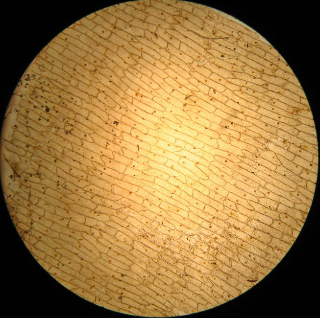
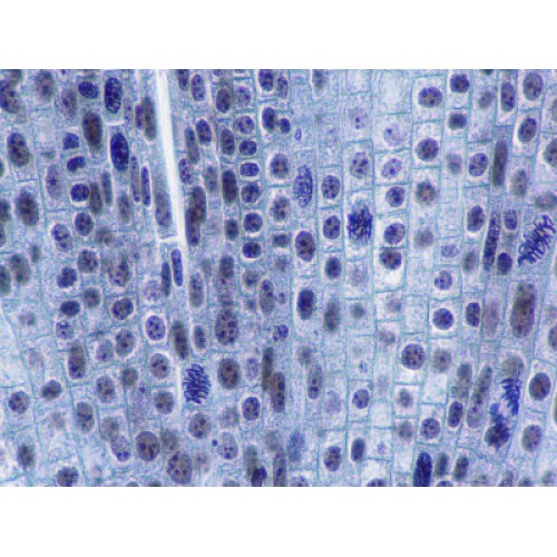
Onion Root Mitosis - Microscopy-UK onion root cells when demonstrating how cell division takes place in Onions have larger chromosomes than most plants and stain dark. The chromosomes are easily observed through a compound light microscope. The cells pictured below are located in the apical meristemof the onion root. The apical

Onion Cell, 400X | Sue Bachus | Flickr
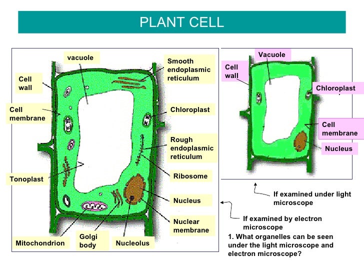
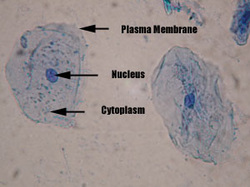
DOC Plant and Animal Cells Microscope Lab 4. Observe the cheek cells under both low and high power of your microscope. Draw a diagram of one cheek cell and label the parts. (You should observe the cell membrane, nucleus, and cytoplasm.) Observation: The following labeled drawings must be completed. Drawings MUST be completed neatly using a pencil/colored pencil. 1. Onion Cell drawing (high power) 2. Cheek cell drawing (any power but preferably high) Drawings, Conclusions and Questions: Onion Cell. Label: Cell Wall. Nucleus

The Microscope
Onion Epidermal Cell Labeled Diagram - schematron.org Labeling of microfilaments in onion bulb scale epidermis cells with. Experimental Preparation of Temporary Mounts of an Onion Peel temporary mount of an onion peel and to record observations and draw labeled diagrams. These cells have a dark stained nucleus and a large vacuole in the centre. Draw a labelled diagram of an onion epidermal cell seen under the microscope. ( 4 marks) e The onion epidermal cells are not green in colour because they lack.

Labelled Diagram Of A Plant Cell Under A Microscope - Micropedia
Observing Cork Cells Under The Microscope » Microscope Club Place the cork dust on the microscope slide with a drop of water, then add another water droplet on top of the cork sample. Cover the prepared slide with a cover slip. Method 2 Alternatively, slice thin cork slices, making sure that ample light can pass through the slice, allowing you to see the cell layout and the individual cells.
Lab #1 microscope structure & function
PDF Onion Cells - Investigation - Exploring Nature 1. Observe the onion tissue under the microscope at 4x, 10x and 40x with lots of light (open diaphragm). 2. Then slowly close the diaphragm while observing the image to find the best light for seeing cellular details. 3. Draw a section of onion skin cells at 10x magnification. 4. Switch to 40x and draw one cell and label it.

Cells & Microscope Activity Unit | Microscope activity, Science cells, Life science activities
Biology Project The Biology Project, an interactive online resource for learning biology developed at The University of Arizona. The Biology Project is fun, richly illustrated, and tested on 1000s of students.
Pictures Of Onion Cells Under A Microscope - Micropedia
Onion Cells Under a Microscope - Requirements/Preparation ... Add a drop of iodine solution on the onion membrane (or methylene blue) Gently lay a microscopic cover slip on the membrane and press it down gently using a needle to remove air bubbles. Touch a blotting paper on one side of the slide to drain excess iodine/water solution, Place the slide on the microscope stage under low power to observe.
Post a Comment for "45 onion cells under microscope with labels"